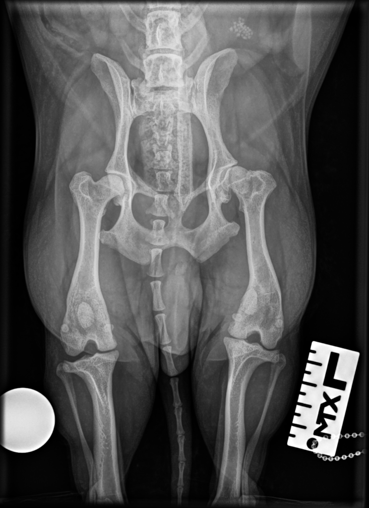
A Hierarchy of Imaging Modalities
Veterinary radiology encompasses a range of imaging techniques of varying cost, availability, and diagnostic power, with conventional X-ray remaining the most widely available modality found in nearly every general practice clinic, while ultrasound, increasingly common even in general practice, provides detailed real-time views of soft tissue and abdominal organs that X-ray cannot capture. Computed tomography and magnetic resonance imaging, offering far greater detail particularly for complex orthopedic, neurological, and oncologic cases, remain concentrated primarily at specialty and referral hospitals due to their substantial equipment cost, reserving these advanced modalities for cases where simpler imaging has proven insufficient.
The Challenge of Patient Stillness
Because diagnostic imaging requires the patient to remain motionless for image acquisition, and animals cannot be verbally instructed to hold still the way human patients can, veterinary imaging frequently requires sedation or full general anesthesia, particularly for advanced modalities like CT and MRI where even small movements can render scans unusable. This anesthesia requirement adds both cost and medical risk to veterinary imaging procedures that human radiology typically avoids, a consideration veterinarians must weigh when determining whether advanced imaging is truly necessary for a given case.
Interpretation and Specialist Collaboration
While general practice veterinarians can interpret straightforward radiographs for common conditions, complex or ambiguous imaging findings are frequently sent to board-certified veterinary radiologists for formal interpretation, a practice that has grown considerably easier with digital imaging and teleradiology services allowing images to be transmitted electronically for remote specialist review. This has extended access to specialist-level imaging interpretation even to smaller clinics without an on-site radiologist, improving diagnostic accuracy for complex cases across a much broader range of veterinary practices than previously possible.
Contrast Studies and Specialized Techniques
Beyond standard imaging, veterinary radiologists employ specialized contrast studies, introducing a contrast agent visible on imaging to highlight specific structures such as the gastrointestinal tract or blood vessels that would otherwise be difficult to distinguish from surrounding tissue on plain imaging. These specialized techniques require additional expertise and equipment but can provide crucial diagnostic information for conditions such as suspected foreign body obstruction or certain vascular abnormalities that plain radiographs alone cannot reliably identify.
Digital Imaging and Teleradiology Growth
The transition from film-based to digital radiography has transformed veterinary imaging workflows, enabling instant image review, easier image sharing with specialists for remote consultation, and elimination of the chemical processing and storage challenges that film-based systems required. This digital transition has particularly benefited smaller and rural veterinary practices, which can now obtain specialist radiologist interpretation through teleradiology services without needing to refer patients physically to a distant specialty hospital for imaging review alone.
Nuclear scintigraphy, a specialized imaging technique using small amounts of radioactive material to highlight areas of abnormal bone metabolism, remains particularly valuable in equine lameness diagnosis for identifying subtle injury sites that conventional radiographs cannot detect.
Radiation safety protocols for veterinary staff performing imaging procedures, including protective lead aprons and positioning techniques that minimize operator exposure, have become standardized practice requirements across accredited veterinary hospitals.
Portable and handheld ultrasound devices have expanded point-of-care imaging capability directly into emergency and ambulatory equine practice settings, allowing rapid assessment without requiring patient transport to a facility with fixed imaging equipment.
Continuing education requirements for veterinary radiology technicians operating imaging equipment help ensure consistent image quality and appropriate safety practice across the profession as imaging technology continues to evolve.
Comparative imaging research across species continues to refine understanding of normal versus abnormal findings, particularly for less commonly imaged species where historical reference data remains comparatively limited.
Equipment maintenance and calibration schedules for imaging machines represent an ongoing operational responsibility for veterinary hospitals, since inconsistent calibration can degrade image quality in ways that compromise diagnostic accuracy over time.
Standardized positioning protocols for common radiographic views help ensure consistent, comparable images across different visits and even across different veterinary facilities evaluating the same patient over time.
Image archiving systems using standardized formats allow veterinary hospitals to maintain searchable historical imaging records, supporting both individual patient care continuity and broader retrospective case review for research purposes.
Mobile CT units, a relatively recent development, have begun extending advanced imaging access to ambulatory equine practice settings previously limited to fixed-facility imaging only.
Image quality assurance reviews conducted periodically help veterinary hospitals identify equipment degradation or technique issues before they meaningfully compromise diagnostic reliability.
Cross-training programs for veterinary technicians in both standard radiography and ultrasound assistance have improved staffing flexibility at practices offering multiple imaging modalities.
Emerging Modalities
Emerging modalities such as elastography, which measures tissue stiffness, are beginning to extend veterinary ultrasound diagnostic capability into new clinical applications.
Standardized reporting templates for common imaging studies help ensure consistent, complete documentation regardless of which staff member performs the interpretation.
Veterinary imaging conferences and journals continue publishing case studies that expand the field’s shared understanding of unusual presentations.
Interdisciplinary imaging rounds, bringing together radiologists, surgeons, and internists to review complex cases together, have improved diagnostic consensus building at larger referral hospitals.
Training and Certification Pathways
Formal certification pathways for veterinary radiology technologists have become more standardized, helping ensure consistent baseline competency across different training programs and employers before staff operate imaging equipment independently on live patients.
Veterinary diagnostic imaging societies maintain accreditation standards that practices can pursue to demonstrate consistent quality to clients and referring veterinarians alike.
Scheduling coordination between imaging departments and the clinicians who depend on their results helps keep overall diagnostic turnaround times reasonable for both routine and urgent cases alike.
Key facts
- Array
Quick checkWhy do many veterinary imaging procedures require sedation or anesthesia?Show answer
Clear diagnostic images require the patient to remain completely still, which most animals cannot be reliably instructed to do, so sedation or anesthesia is often used to prevent motion blur and allow proper positioning.
Sources
- Array
Editorially reviewed by Specialty Digest Editorial TeamLast reviewed November 4, 2026Researched and drafted with AI assistanceReport an issue