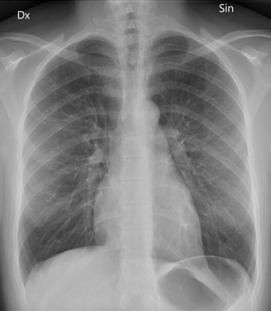
X-ray imaging is a medical and diagnostic technique that sends X-rays, a form of ionizing electromagnetic radiation, through the body and records how much of the beam each tissue absorbs. Dense structures such as bone appear white and softer tissue appears in shades of gray, which makes the method especially useful for finding fractures and many other problems inside the body.
History of X-ray imaging
The technology began in 1895, when the German physicist Wilhelm Conrad Roentgen was studying cathode radiation, which occurs when an electrical charge is applied across two metal plates inside a glass tube holding rarefied gas. Although his apparatus was screened off, he noticed a faint light on light-sensitive screens nearby. Further tests showed that the glow was caused by a penetrating, previously unknown type of radiation.
That discovery earned Roentgen the first Nobel Prize in Physics in 1901, awarded for the remarkable rays that were later named after him. The Nobel Foundation notes that X-ray radiation soon became a powerful tool both for physical experiments and for examining the inside of the body.
The next major step came in the 1960s with computed tomography, which turned X-ray measurements into cross-sectional images. The FDA describes the discovery of X-rays and the invention of CT as major advances in medicine.
How X-ray imaging works
Every X-ray examination follows the same basic principle. A beam is passed through the body, where part of it is absorbed or scattered by internal structures, and the remaining X-rays reach a detector. The detector can be film or a digital sensor, and its signal is recorded or processed by a computer.
Different tissues absorb radiation to different degrees. MedlinePlus explains that bones absorb the most and therefore appear white, fat and other soft tissues absorb less and appear gray, and air absorbs the least, so the lungs look black. A specialist called a radiologist reviews the images and reports the findings to the health care provider who ordered them.
X-rays are called ionizing radiation because they carry enough energy to knock electrons out of atoms. According to the FDA, that energy can potentially damage DNA, which is why radiation doses in medical imaging are measured and regulated.
Types of X-ray imaging
The FDA groups the main X-ray based methods into radiography, fluoroscopy and computed tomography. Radiography, often called a conventional X-ray, records a single image for later evaluation. Mammography is a special type of radiography used to image the internal structures of the breast.
Fluoroscopy displays a continuous X-ray image on a monitor, so clinicians can watch a procedure or follow a contrast agent, commonly called dye, as it moves through the body. Computed tomography records many X-ray images as the detector moves around the patient, and a computer reconstructs them into cross-sectional slices.
MedlinePlus lists many specific examinations built on these methods, including chest, abdominal, bone and dental X-rays and bone density scans. Some use a contrast material to improve visualization of particular organs or vessels.
Applications of X-ray imaging
The most familiar use is checking for fractures. Chest X-rays can detect pneumonia, and mammograms use X-rays to look for breast cancer. X-rays are also built into other tests, such as CT scans.
The FDA lists several broader roles. X-ray imaging helps diagnose disease and monitor therapy without surgery, supports medical and surgical treatment planning, and guides clinicians as they place catheters or stents, treat tumors or remove blood clots and other blockages.
An X-ray is often the first image taken, and the result may settle the question on its own or lead to a further test, such as computed tomography or MRI, is needed.
Advantages and disadvantages of X-ray imaging
The FDA calls X-ray exams a valuable medical tool that is noninvasive and painless. MedlinePlus adds that in most cases little or no preparation is needed.
The main drawback is radiation exposure. The FDA states that the risk of developing cancer from medical imaging radiation is generally very small, but that the lifetime risk rises with the dose and with the number of exams. It also depends on the age of the person at exposure, with younger patients at larger risk, on sex, with women at a somewhat higher risk, and on which body region is exposed.
For scale, MedlinePlus says the radiation from a chest X-ray is about the same as the natural background radiation a person receives over roughly 10 days. Fluoroscopy can involve relatively high doses during long, complex procedures, and tissue effects such as skin reddening, hair loss or cataracts occur only at relatively high levels of exposure and are rare for most types of imaging.
A second limit is the picture itself. A radiograph compresses a three-dimensional body into one flat image, so structures can overlap, and soft tissues show less contrast than bone. Contrast agents carry their own small risk of reactions.
X-ray imaging vs. CT
Radiography and CT use the same kind of radiation but answer different questions. A conventional X-ray produces one projection, while a CT scanner combines many projections into slices and three-dimensional views that show organs, blood vessels and soft tissue in more detail.
That detail has a cost in dose. The FDA says a CT exam involves a higher radiation dose than conventional radiography because the image is reconstructed from many individual projections. Where an examination can be answered without ionizing radiation, the FDA notes that ultrasound or MRI may be considered, which is why the choice between methods is made case by case by clinicians. This entry is general background and not medical advice.
More in Medicine
Key facts
- Radiography, fluoroscopy and CT all pass an X-ray beam through the body to a detector, where part of the beam has been absorbed or scattered.1
- Roentgen noticed the new penetrating radiation in 1895 and received the Nobel Prize in Physics in 1901.3
- On X-ray images bones appear white, soft tissues gray and air black.2
- Mammography is a special type of radiography, and fluoroscopy shows a continuous image in real time.1
- The FDA says ionizing radiation can potentially damage DNA and may elevate lifetime cancer risk, though the risk from imaging is generally very small.1
- A chest X-ray delivers about the radiation of roughly 10 days of natural background exposure.2
This entry explains a medical term. It is not medical advice. If you are concerned about your health, speak with a qualified health professional.
Related concepts
Quick checkWhy do bones look white on an X-ray?Show answer
Bones absorb the most radiation, so little reaches the detector behind them and they appear white.
Sources
- 1. U.S. Food and Drug Administration, Medical X-ray Imaging, https://www.fda.gov/radiation-emitting-products/medical-imaging/medical-x-ray-imaging
- 2. MedlinePlus (U.S. National Library of Medicine), X-Rays, https://medlineplus.gov/xrays.html
- 3. The Nobel Prize, Wilhelm Conrad Roentgen Facts, Physics 1901, https://www.nobelprize.org/prizes/physics/1901/rontgen/facts/
Editorially reviewed by Specialty Digest Editorial TeamLast reviewed November 4, 2026Researched and drafted with AI assistanceReport an issue