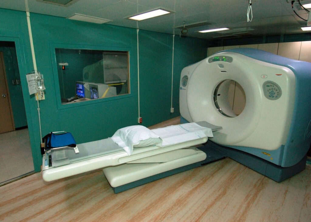
Computed tomography, or CT, is a medical imaging technique that uses a rotating X-ray source and a computer to produce cross-sectional pictures of the body. Each image is a thin slice, and slices can be combined into three-dimensional views of organs, bones, blood vessels and other tissues.
History of computed tomography
CT grew out of X-ray imaging, which showed the inside of the body but flattened it into a single picture. During the 1960s, the British engineer Godfrey Hounsfield, who worked at the Central Research Laboratories of the company EMI in London, developed an apparatus that sent clusters of X-ray beams through the body from different angles and registered what emerged. Advanced computer calculations then turned those measurements into images of different cross sections of the body, and the same data could also form the basis of three-dimensional images.
The physicist Allan Cormack shared the Nobel Prize in Physiology or Medicine with Hounsfield in 1979. The prize was given for the development of computer assisted tomography, as the technique was then called.
The names used for the method have varied. The FDA notes that CT is sometimes called computerized tomography or computed axial tomography, and the abbreviation CAT scan is still widely used in everyday language.
How computed tomography works
A CT system is built around a circular opening, often described as donut-shaped. A motorized table moves the patient through the opening while an X-ray source and a detector assembly rotate around the body. According to the FDA, a single rotation typically takes a second or less.
During each rotation the source produces a narrow, fan-shaped beam that passes through a section of the body. Rows of detectors on the opposite side register the X-rays that get through, and each reading is a snapshot taken from one angle. Many snapshots are collected in a full rotation.
The image is not recorded directly. A computer reconstructs the individual snapshots into one or more cross-sectional slices, which the FDA compares to the slices of a loaf of bread. CT images of internal organs, bones, soft tissue and blood vessels show more clarity and detail than conventional X-ray images such as a chest radiograph.
MedlinePlus adds that a scan usually takes a few minutes, although some last up to 30 minutes depending on the area examined. The test is painless, the machine makes whirring sounds, and the person may be asked to hold their breath for short periods.
Types of CT scans
The FDA says CT scans can be performed on every region of the body and for several kinds of purpose: diagnostic, treatment planning, interventional and screening. Diagnostic scans look for the cause of symptoms. Interventional scans guide a procedure as it happens, for example when a physician needs to find the best place to take a biopsy sample.
Scans also differ in whether a contrast material is used. Contrast helps certain parts of the body show up more clearly, and MedlinePlus says it can be given as a liquid to swallow, as an injection or as an enema, depending on the area being examined.
Applications of computed tomography
MedlinePlus lists common reasons for CT scans, including fractures, cancer, tumors or masses, blood clots, signs of heart disease, spinal conditions and lung disease. Scans may also be used to check for internal bleeding or injury after trauma, to evaluate the brain for certain conditions and to guide some treatments.
The FDA groups the uses into three roles. CT helps a physician diagnose disease, trauma or abnormality, plan and guide interventional or therapeutic procedures, and monitor the effectiveness of therapy, such as cancer treatment. Most scans are done as outpatient procedures.
Animals are imaged with related methods, which are covered in the entry on veterinary radiology.
Advantages and disadvantages of computed tomography
The FDA states that, when used appropriately, the benefits of a CT scan far exceed the risks. The detailed images can provide the information needed to diagnose and plan treatment for many conditions in adults and children, and in some cases they may remove the need for exploratory surgery.
The main concern is radiation. CT uses ionizing radiation at levels higher than a standard X-ray, because the image is built from many projections, although the dose is kept as low as possible. The FDA says the exposure may cause a small increase in a person’s lifetime risk of developing cancer.
Children need particular attention. The FDA explains that the cancer risk per unit of radiation dose is higher for younger patients, who also have more years in which effects could appear. It adds that exposure settings designed for adults can deliver a larger dose than necessary to a child, so settings need to be adjusted for pediatric patients.
A second concern is contrast material, which can occasionally cause reactions. The FDA also states that, at this time, it does not see a benefit to whole-body scanning of individuals who have no symptoms.
CT vs. MRI
CT and MRI both produce cross-sectional images, but they rely on different physics. CT measures how X-rays are absorbed along many paths through the body. MRI uses strong magnetic fields and radio waves instead of ionizing radiation.
The FDA tells referring physicians to consider examinations that use less or no radiation, such as ultrasound or MRI, when they can answer the clinical question. Because each method shows some tissues better than others, the choice depends on what must be seen, and this entry is general background rather than medical advice.
More in Medicine
Key facts
- CT uses specialized X-ray equipment to produce cross-sectional images, or slices, of the body.1
- In a CT system the X-ray source and detectors rotate around the patient, and a single rotation typically takes a second or less.1
- Godfrey Hounsfield and Allan Cormack shared the 1979 Nobel Prize in Physiology or Medicine for computer assisted tomography.3
- CT uses ionizing radiation at levels higher than a standard X-ray.2
- The FDA does not see a benefit to whole-body CT scanning of individuals without symptoms.1
- A CT scan usually takes a few minutes, though some last up to 30 minutes.2
This entry explains a medical term. It is not medical advice. If you are concerned about your health, speak with a qualified health professional.
Related concepts
Quick checkWho shared the 1979 Nobel Prize for computer assisted tomography?Show answer
Godfrey Hounsfield and Allan Cormack.
Sources
- 1. U.S. Food and Drug Administration, Computed Tomography (CT), https://www.fda.gov/radiation-emitting-products/medical-x-ray-imaging/computed-tomography-ct
- 2. MedlinePlus (U.S. National Library of Medicine), CT Scans, https://medlineplus.gov/ctscans.html
- 3. The Nobel Prize, Godfrey N. Hounsfield Facts and The Nobel Prize in Physiology or Medicine 1979, https://www.nobelprize.org/prizes/medicine/1979/hounsfield/facts/
- 4. U.S. Food and Drug Administration, Medical X-ray Imaging, https://www.fda.gov/radiation-emitting-products/medical-imaging/medical-x-ray-imaging
Editorially reviewed by Specialty Digest Editorial TeamLast reviewed November 4, 2026Researched and drafted with AI assistanceReport an issue