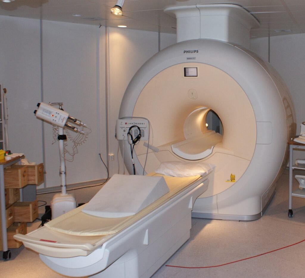
Magnetic resonance imaging, or MRI, is a medical imaging technology that uses strong magnetic fields and radio waves to make detailed pictures of the inside of the body. The U.S. Food and Drug Administration describes it as a procedure for imaging internal structures. Unlike X-ray based scans, it builds images from the behavior of atomic nuclei, mainly the nuclei of hydrogen in water, inside a magnetic field. It is especially useful for soft tissues such as the brain, muscles and joints.
History of MRI technology
The physics came first. The Nobel Assembly at Karolinska Institutet explains that atomic nuclei in a strong magnetic field rotate at a frequency that depends on the strength of the field. They can absorb radio waves of the same frequency, an effect called resonance, and emit radio waves when they return to their earlier energy level. Those discoveries were awarded the Nobel Prize in Physics in 1952, and for decades afterward magnetic resonance was used mainly to study the chemical structure of substances.
In the early 1970s two researchers turned the effect into an imaging method. Paul Lauterbur of Urbana, Illinois, discovered that adding gradients to the magnetic field made two-dimensional pictures possible, because analyzing the emitted radio waves revealed where they came from. In 1973 he described how adding gradient magnets to the main magnet could show a cross section of tubes of ordinary water surrounded by heavy water. Peter Mansfield of Nottingham, England, developed the use of gradients further and showed that extremely fast imaging could be achieved. The two shared the 2003 Nobel Prize in Physiology or Medicine.
How MRI technology works
Water explains why the method suits the body. The Nobel committee points out that water makes up about two thirds of human body weight and that the hydrogen nuclei in a water molecule act like microscopic compass needles. In a strong magnetic field they line up with it. Radio pulses then disturb that alignment, and as the nuclei settle back they give off signals that the scanner detects.
Tissues differ in water content and in how freely water moves, and many diseases change the water content, so the signals produce contrast between tissues. Gradient magnets, the idea Lauterbur introduced, mark where each signal comes from. Computer processing turns the signals into two-dimensional slices or three-dimensional images.
The FDA describes the hardware. An electric current passed through coiled wires creates a temporary magnetic field in the patient’s body, and a transmitter and receiver in the machine send and detect radio waves. For some examinations, gadolinium-based contrast agents are usually given through a vein in the arm to change the contrast of the image.
Types of MRI
Most scans are structural, showing the shape and condition of tissues and organs. Functional MRI measures changes in blood flow linked to brain activity and is widely used in neuroscience research. Magnetic resonance angiography focuses on blood vessels. Scans with contrast agents highlight tissues that take up the agent differently. By changing the timing of the radio pulses, operators can emphasize different tissue properties, which is why one examination often includes several image sets.
Operators also vary the strength of the magnet. Higher-field scanners can capture finer detail but require stricter safety controls.
Applications of MRI technology
The National Health Service in England says an MRI scan helps diagnose conditions, plan treatment and check how well treatment is working. It can be used to find the cause of symptoms, to examine bones and organs, and to track the size of a tumor during and after cancer treatment.
A scan takes place in a hospital and is run by a radiographer from a separate room. The machine is a large unit with a short tunnel and a flat bed that slides in. Patients lie still, wear ear plugs or headphones because the scanner makes loud bangs and clicks, and may be asked to hold their breath briefly. The NHS gives a typical duration of 15 minutes to 1 hour, while the FDA says 20 to 90 minutes depending on the body part. A radiologist examines the images and reports to the referring doctor, and the NHS says results usually take one to two weeks.
Advantages and disadvantages of MRI technology
The main advantage is detail. MRI produces sharp images of soft tissue using magnetic fields and radio waves instead of X-rays, and it can image in any plane without moving the patient.
The drawbacks are practical and safety-related. Scans are long, noisy and take place in an enclosed space, which the NHS notes can be distressing for people with claustrophobia. Because the magnet is strong, metal is a concern. The NHS says patients complete a screening form about pregnancy, earlier surgery, kidney problems, allergies and metal in the body such as surgical clips, pacemakers and dental work, and are asked to remove metal items. The FDA has also issued a safety communication on implantable infusion pumps in the MR environment. Contrast agents can cause side effects, which the NHS describes as usually mild and short-lived, such as nausea, a rash, a headache or a metallic taste. This entry is general background, and decisions about any scan rest with the patient and their clinicians.
MRI vs. CT
Computed tomography, or CT, uses X-rays and a computer to build cross-sectional images, while MRI uses magnetic fields and radio waves. CT scans are usually faster, which can matter in emergencies. MRI usually shows greater contrast between soft tissues. Which test is used depends on the clinical question, the part of the body and the patient’s circumstances, and the choice is made by clinicians.
More in Medicine
Key facts
- MRI scanners use strong magnetic fields and radio waves to make images of internal body structures.3
- Paul Lauterbur and Peter Mansfield shared the 2003 Nobel Prize in Physiology or Medicine.1
- In 1973, Lauterbur described gradient magnets that showed a cross section of tubes of ordinary water surrounded by heavy water.1
- Magnetic resonance work earned the Nobel Prize in Physics in 1952.1
- Water makes up about two thirds of human body weight, which is why MRI suits medical imaging.1
- The NHS gives a typical MRI scan time of 15 minutes to 1 hour.2
- The FDA says a typical MRI scan lasts 20 to 90 minutes, depending on the body part.3
This entry explains a medical term. It is not medical advice. If you are concerned about your health, speak with a qualified health professional.
Related concepts
In the news
Quick checkWho won the 2003 Nobel Prize for discoveries behind MRI?Show answer
Paul Lauterbur and Peter Mansfield, for discoveries that led to modern magnetic resonance imaging.
Sources
- 1. The Nobel Prize, The Nobel Prize in Physiology or Medicine 2003 press release, https://www.nobelprize.org/prizes/medicine/2003/press-release/
- 2. National Health Service (England), MRI scan, https://www.nhs.uk/conditions/mri-scan/
- 3. U.S. Food and Drug Administration, MRI (Magnetic Resonance Imaging), https://www.fda.gov/radiation-emitting-products/medical-imaging/mri-magnetic-resonance-imaging
Editorially reviewed by Specialty Digest Editorial TeamLast reviewed November 4, 2026Researched and drafted with AI assistanceReport an issue