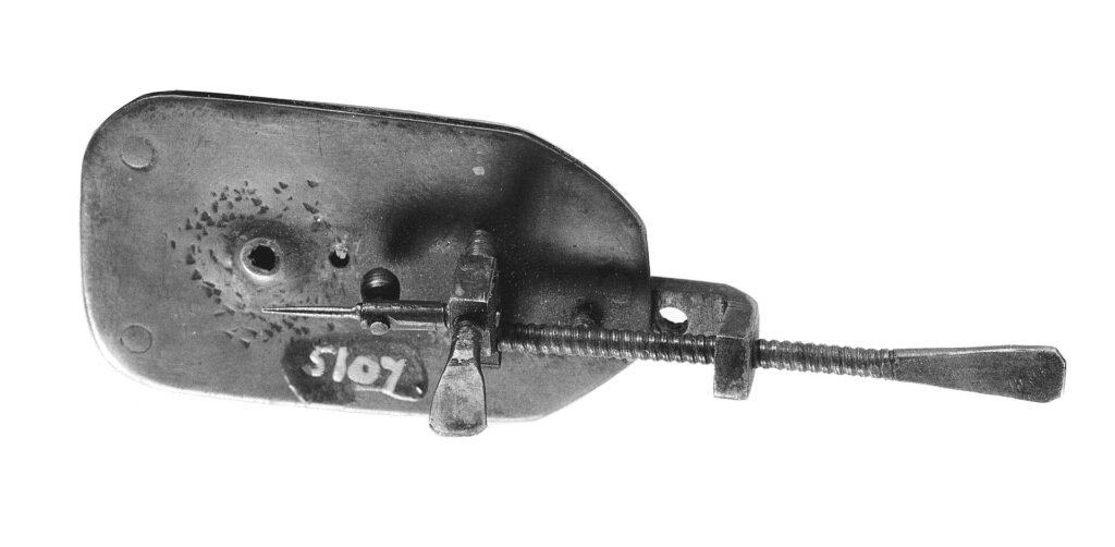
A microscope is an instrument that magnifies objects too small to see with the naked eye. The first compound microscopes appeared in the Netherlands around 1590, Robert Hooke used one to describe cells in 1665, and electron microscopes developed from the early 1930s extended the view to the scale of molecules.
History of the microscope
A review of 300 years of light microscopy reports that Hans and Zacharias Janssen made a microscope with lenses in a tube around 1590, although no observations made with it were published. Galileo built a compound microscope in 1609, and Giovanni Faber coined the word microscope in 1625.
The instrument became a scientific tool in the 17th century. Robert Hooke published Micrographia in 1665 and described the tiny pores in cork as cells. Antonie van Leeuwenhoek, who lived from 1632 to 1723, built more than 500 simple one-lens microscopes and reported what he saw to the Royal Society in a series of letters.
Advances in optics came next. In 1883 Ernst Abbe and Otto Schott’s high-quality lenses made diffraction-limited microscopes possible. Stains such as Gram’s method in 1884 made bacteria and tissues easier to tell apart, and in the 20th century phase contrast, fluorescence and confocal methods added new ways to see transparent or labeled structures.
Electron microscopy grew from work in Berlin. Ernst Ruska learned of Hans Busch’s idea that a magnetic coil can focus a beam of electrons like a glass lens focuses light. In a thesis submitted in May 1929 Ruska calculated and tested the imaging properties of such a coil and obtained the first recorded electron-optical images. He shared the 1986 Nobel Prize in Physics for this work, together with Gerd Binnig and Heinrich Rohrer.
Who invented the microscope?
The invention cannot be assigned to one person. The Janssens are usually credited with the first compound microscope, but the evidence is thin because no observations from their instrument were published. Galileo made a compound microscope too, and many craftsmen working with lenses in the Netherlands and Italy were exploring similar ideas.
What is clearer is who turned the microscope into a tool of discovery. Hooke recorded detailed drawings in Micrographia, and Leeuwenhoek’s letters described tiny living organisms that nobody had seen before.
How a microscope works
A light microscope uses two sets of lenses. The first lens, the objective, receives light from the illuminated specimen and forms a real image, and the second lens, the ocular or eyepiece, magnifies that image. Together they can enlarge an object up to about 1,000 times.
Resolution, not magnification, sets the practical limit. As described in the review, the Abbe limit is roughly half the wavelength of the light used, so details smaller than that blur together, no matter how much the image is enlarged.
Electron microscopes avoid this limit because electrons have a much shorter wavelength than visible light. Magnetic coils act as lenses, and the electron beam produces images at a far finer scale, though specimens must be prepared and examined in a vacuum.
Seeing detail also depends on contrast, because most cells are nearly transparent. Joseph von Gerlach showed in 1858 that carmine dye stains cell nuclei differently from their surroundings, Camillo Golgi introduced silver staining in 1873, and Hans Christian Gram invented his staining method in 1884. Fluorescein, the first fluorescent stain, followed in 1871.
Fluorescent labels advanced in the late 20th century. Green fluorescent protein was first isolated from the jellyfish Aequorea victoria in 1962, and in 1994 Martin Chalfie and colleagues showed that it could glow in other organisms, which made it possible to tag specific proteins.
Types of microscopes
Common optical types include the compound brightfield microscope, the stereo microscope for dissection and inspection, and phase contrast and differential interference contrast systems, which show transparent cells without stains. Fritz Zernike’s phase contrast method earned a Nobel Prize in 1953.
Fluorescence microscopes make labeled structures glow. Confocal microscopes, patented by Marvin Minsky in 1961, record thin optical slices, and super-resolution methods such as STED, proposed in 1994 and demonstrated in 1999, and PALM and STORM, introduced in 2006, get below the old limit. Scanning tunneling microscopes, from the work honored in 1986, image atoms on surfaces.
Advantages and disadvantages of light and electron microscopes
Light microscopes are comparatively cheap, easy to use and able to show living cells in color, but their resolution is limited by the wavelength of light. Electron microscopes show far finer detail, yet they are expensive, need specially prepared specimens and generally cannot image living material because the sample sits in a vacuum.
Choosing between them depends on the question. A school laboratory needs a light microscope, while a researcher studying the shape of a virus needs electron or super-resolution methods.
Impact of the microscope
The microscope made cell biology, microbiology and pathology possible. Seeing cells, bacteria and tissue structure changed how medicine understood disease, and later tools such as green fluorescent protein, honored with a Nobel Prize in Chemistry in 2008, let researchers follow molecules inside living cells.
Beyond biology, microscopes now inspect metals, minerals and manufactured parts. They also tie into neuroscience, where staining and imaging reveal how a neuron connects with others.
Uses of the microscope today
Hospital laboratories use microscopes to examine blood and tissue samples, and research labs use them to study cells, materials and nanostructures. Schools use simple compound microscopes to teach biology, and engineers use electron and optical microscopes to inspect microchips and coatings.
FAQs about the microscope
How strong can a light microscope be? A typical compound microscope enlarges up to about 1,000 times, and the useful limit is set by resolution rather than magnification.
What is the difference between a light and an electron microscope? A light microscope focuses visible light with glass lenses. An electron microscope focuses a beam of electrons with magnetic fields and can show much finer detail.
Who first saw cells? Robert Hooke named them in 1665 when he looked at thin slices of cork, which showed small box-like pores.
More in Biology
Key facts
- Array
Related concepts
In the news
Quick checkWho invented the microscope?Show answer
No one person can be named. Hans and Zacharias Janssen are usually credited with a compound microscope around 1590, Galileo made one in 1609, and Hooke and Leeuwenhoek made it a scientific tool.
Sources
- Array
Editorially reviewed by Specialty Digest Editorial TeamLast reviewed November 4, 2026Researched and drafted with AI assistanceReport an issue