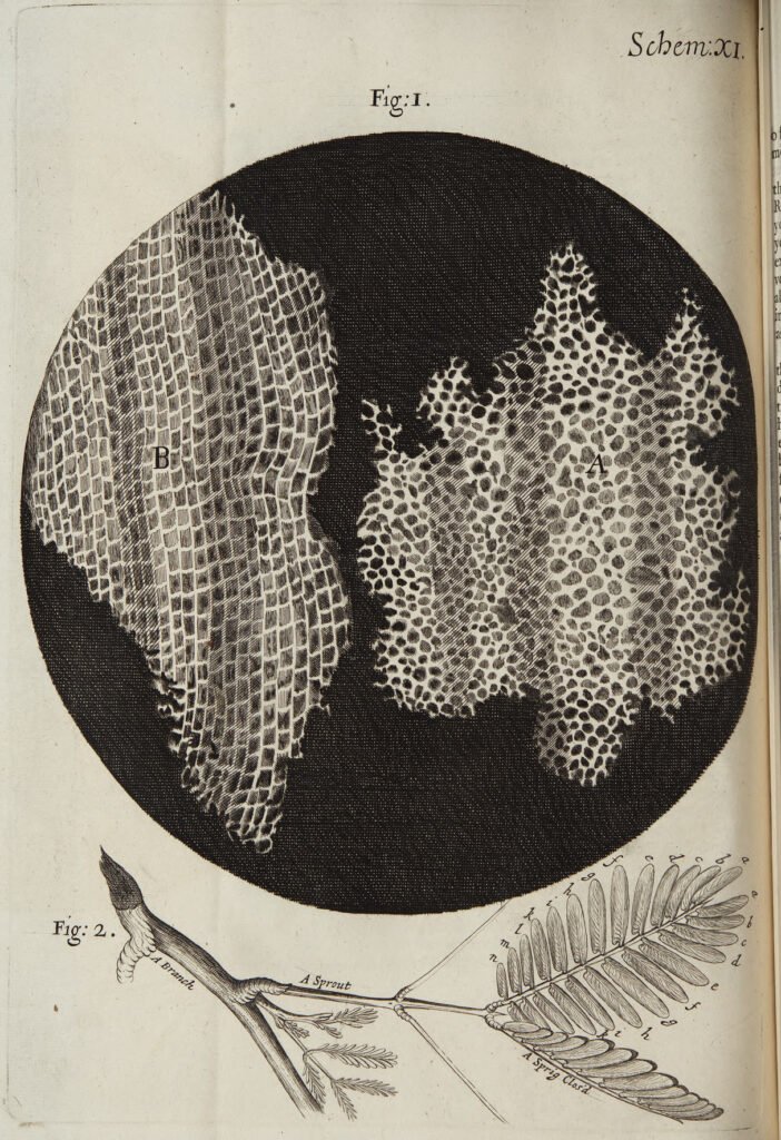
The discovery of cells began in 1665, when the English scientist Robert Hooke examined a thin slice of cork under a microscope and described its honeycomb-like chambers as “cells.” A few years later the Dutch merchant Antony van Leeuwenhoek became the first person to observe living single-celled organisms, which he called animalcules. Their work opened the way to cell theory, the idea that all living things are made of cells.
Background
By the 1660s, magnifying lenses were old technology, but seeing small things clearly was still difficult. Compound microscopes, which use more than one lens, had been invented around 1595. Early versions could not usefully magnify objects more than about twenty or thirty times, so they revealed little of the hidden structure of living things.
Progress came from two very different men. Robert Hooke was born in 1635 on the Isle of Wight and trained under the chemist Robert Boyle at Oxford. In 1662 he became Curator of Experiments of the newly formed Royal Society of London, which meant he demonstrated new experiments at its weekly meetings. He built a compound microscope with its own lighting system and used it to study insects, sponges, feathers and plants.
Antony van Leeuwenhoek was a cloth merchant in Delft, in the Netherlands. He had no university education and knew only Dutch, yet he taught himself to grind lenses and became one of the great observers of his century.
How cells were discovered
Hooke and the cork cells
Hooke published his findings in Micrographia in 1665. The book was richly illustrated and became a best seller. The diarist Samuel Pepys stayed up until 2:00 in the morning reading it and called it “the most ingenious book that I ever read in my life.”
In Observation XVIII, Hooke lit a thin piece of cork with a lens and saw that it was “all perforated and porous, much like a Honey-comb.” He wrote that these pores, or cells, were the first microscopical pores he had ever seen, and perhaps the first anyone had seen. The University of California Museum of Paleontology notes that the box-like chambers reminded him of the cells of a monastery. He found similar structures in wood and other plants.
What Hooke saw, however, were the empty walls of dead plant tissue. He did not know that living cells fill such spaces, but his word for them stuck.
Leeuwenhoek and the first living cells
Leeuwenhoek was born in Delft on 24 October 1632. Before 1668 he learned to grind lenses and began building simple microscopes, probably after seeing a copy of Micrographia. Each instrument used a single tiny lens set in a brass plate. He made more than 500, and fewer than ten survive. With careful lighting, they magnified more than 200 times and gave clearer images than the compound microscopes of his day.
In 1673 he began writing letters to the Royal Society, and he kept doing so for fifty years. The letters were translated and printed in the society’s Philosophical Transactions. In the 1670s he saw bacteria and protozoa, which he called animalcules. A letter of 1674 described lake water containing green spirals that modern readers recognize as the alga Spirogyra. He also described bacteria taken from scrapings of his own teeth, along with blood cells and sperm cells.
To check these surprising reports, the Royal Society asked Hooke in 1678 to repeat the observations. Hooke confirmed them, although he complained that Leeuwenhoek’s single-lens microscopes strained his eyes. Leeuwenhoek was elected a full member of the society in 1680.
Hooke vs. Leeuwenhoek
The two men are often blended into one story, but they saw different things. Hooke used a compound microscope and examined dead plant material, so he recorded the walls of cells rather than the living contents. Leeuwenhoek used single-lens instruments and examined water, blood and other fluids, so he met living cells in motion. Hooke named the structure, and Leeuwenhoek showed that whole organisms can consist of a single cell.
Leeuwenhoek is sometimes called the inventor of the microscope. He was not. Compound microscopes existed decades before he was born. His skill lay in making unusually good lenses and in describing exactly what he saw, often with drawings by a hired illustrator.
Impact of the discovery
Later advances in lenses, microscope construction and staining techniques let scientists see structures inside cells. By the late 1830s the botanist Matthias Schleiden and the zoologist Theodor Schwann were studying tissues and proposed the unified cell theory. It holds that one or more cells make up all living things, that the cell is the basic unit of life, and that new cells arise from existing cells. Rudolf Virchow later made important contributions to it.
The last idea explains how organisms grow and repair themselves, as in mitosis, the division of one cell into two. It also underlies modern cell biology, the study of how cells are built and function. Leeuwenhoek’s microorganisms opened the field of microbiology and showed that most life on Earth is invisible to the naked eye.
The tools have changed greatly. A modern microscope can reveal structures far smaller than anything Hooke or Leeuwenhoek could resolve, and in medicine, cytotechnologists stain and examine samples of cells to look for signs of disease. Both pioneers worked in London or corresponded with it through the Royal Society, and their story shows how a shared scientific community helped turn individual observations into accepted knowledge.
FAQs
Who discovered cells? Robert Hooke first described and named cells in 1665, using cork. Antony van Leeuwenhoek first observed living single-celled organisms in the 1670s.
Why are they called cells? Hooke chose the word because the small, box-like compartments in cork looked like the cells of a monastery.
What did Hooke actually see in cork? He saw the cell walls of dead plant tissue, which formed a pattern of small empty chambers.
Did Leeuwenhoek invent the microscope? No. Compound microscopes were invented around 1595. Leeuwenhoek made exceptional single-lens microscopes and used them to make major discoveries.
More in Biology
Key facts
- Robert Hooke published Micrographia in 1665 and described the cell-like pores of cork, which he called cells.12
- Antony van Leeuwenhoek began writing to the Royal Society in 1673 and observed bacteria and protozoa in the 1670s.13
- The Royal Society asked Hooke in 1678 to confirm Leeuwenhoek's observations, and he did.1
- Leeuwenhoek's microscopes had a single lens and magnified more than 200 times.1
- Schleiden and Schwann proposed the unified cell theory in the late 1830s.4
Quick checkWho discovered cells?Show answer
Robert Hooke named cells in 1665 after viewing cork under a microscope, and Antony van Leeuwenhoek first saw living single-celled organisms in the 1670s.
Sources
- University of California Museum of Paleontology, Robert Hooke, https://ucmp.berkeley.edu/history/hooke.html
- University of California Museum of Paleontology, Antony van Leeuwenhoek, https://ucmp.berkeley.edu/history/leeuwenhoek.html
- Molecular Expressions, Florida State University, Antonie van Leeuwenhoek, https://micro.magnet.fsu.edu/optics/timeline/people/leeuwenhoek.html
- OpenStax, Biology 2e, 4.1 Studying Cells, https://openstax.org/books/biology-2e/pages/4-1-studying-cells
- Robert Hooke, Micrographia (1665), Project Gutenberg edition, https://www.gutenberg.org/files/15491/15491-h/15491-h.htm
Editorially reviewed by Specialty Digest Editorial TeamLast reviewed November 4, 2026Researched and drafted with AI assistanceReport an issue