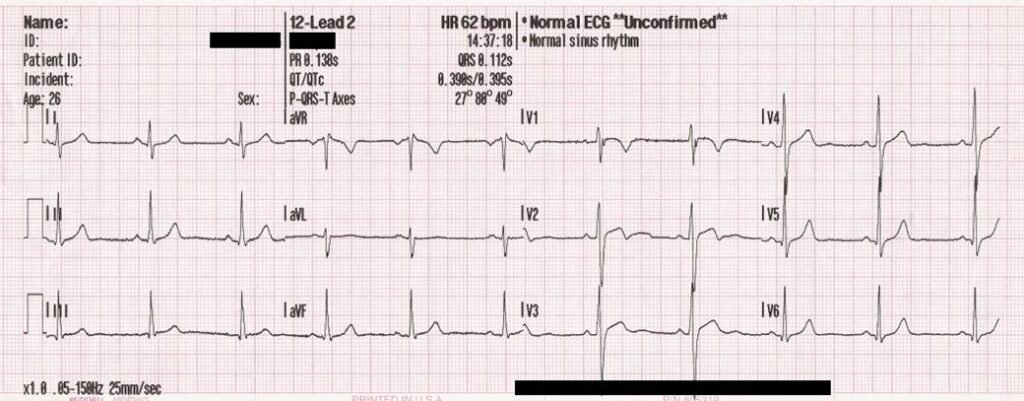
An electrocardiogram (ECG, also written EKG) is a test that records the electrical signals the heart makes each time it beats. MedlinePlus, the health information service of the U.S. National Library of Medicine, describes it as a simple, painless and quick test. The recording appears as wavy lines with sharp spikes, and a trained clinician reads the pattern to judge heart rate, rhythm and the timing of the signals that move through the heart. The device that makes the recording is an electrocardiograph, and the instrument built by Willem Einthoven in the early 1900s made the test practical.
Background: history of the electrocardiogram
The idea rests on a discovery of the 1800s: each heartbeat produces weak electrical currents that can be detected on the body’s surface. Early recordings used a capillary electrometer, an instrument invented in the 1870s by Gabriel Lippmann in which a column of mercury moves as the electrical potential changes. In 1887 the British physiologist Augustus Waller used one to make the first recording of a human electrocardiogram, with electrodes placed on the chest.
Waller’s device was slow and imprecise, and he did not see its clinical value. In 1911 he still doubted that the technique would find extensive use in hospitals. Willem Einthoven, a physiology professor at Leiden University in the Netherlands, took a different view. According to IEEE Spectrum, he began analyzing Waller’s data in 1901, found errors in the visual recordings and sought help from the physicist Hendrik Lorentz to correct them.
Einthoven then built a far more sensitive instrument, the string galvanometer. It used a very thin conducting filament stretched between strong electromagnets. When current from the heart passed through the filament, it moved, and a beam of light shining past it recorded the motion on photographic paper. The same type of device had earlier been used to detect weak signals on long-distance cables, which linked it to the telegraph era.
Sources date the working instrument anywhere from 1901 to 1905, depending on whether they count the first prototype, the practical version or the first published recordings. The Nobel Prize organization dates its construction to 1903. The first machine was far from convenient: one historical account says it weighed about 600 pounds, needed five people to operate and required the patient to place a leg and an arm in buckets of water.
Einthoven also gave the tracing its vocabulary. He named the deflections P, QRS and T, and the imaginary triangle used to describe the standard limb connections is called Einthoven’s triangle. In 1924 he received the Nobel Prize in Physiology or Medicine for his discovery of the mechanism of the electrocardiogram. His string galvanometer later received an IEEE Milestone.
How an electrocardiogram works
The heart’s four chambers contract in a set rhythm because electrical signals tell them to. Those signals spread to the skin, where electrodes, which are small sensors that stick to the skin, can pick them up. During a standard test, MedlinePlus explains, a provider places several electrodes on the arms, legs and chest, and wires connect them to a computer or a dedicated ECG machine. The person lies still while the activity is recorded on a screen or printed on paper. The whole test takes only a few minutes.
Modern machines replaced the photographic paper, the cooling water and the buckets with sticky electrodes and digital recording, but the principle is the one Einthoven used: measure tiny voltage changes between points on the body over time.
What an ECG shows
MedlinePlus breaks the tracing into three main parts. The P wave reflects how well the upper chambers of the heart work. The QRS complex, the sharp spike, reflects the lower chambers. The T wave shows how well the heart recovers after the chambers contract.
From these shapes a provider can see how fast the heart is beating, whether the rhythm is steady or irregular, and the strength and timing of the signals passing through each part of the heart. Sometimes the tracing also helps estimate the size and position of the chambers.
Types of ECG tests
A resting ECG is the most familiar kind and may be done in an office, an outpatient clinic or a hospital before surgery. An ECG can also be part of a stress test, which records the heart while it works harder. Ambulatory electrocardiography records the heart for a longer period while a person goes about daily life, using devices such as Holter monitors and event monitors. Which test suits a person is a decision for a health care provider.
Uses and limits of the ECG
Clinicians use the test to look for abnormal heart activity. MedlinePlus says the results cannot diagnose a heart condition on their own. Combined with other tests and a medical history, however, they help providers diagnose and monitor conditions such as arrhythmia, cardiomyopathy, coronary artery disease, heart attack, heart failure, heart valve disease and congenital heart defects.
Providers may also use an ECG to check whether the heart is healthy enough for surgery, to see how well a new heart medicine is working, or to follow a patient after a pacemaker is implanted. The tracing is also what lets a defibrillator recognize a rhythm that needs a shock.
The test is mainly used for people who have symptoms or a known heart condition. MedlinePlus notes that it is not generally used to screen people without symptoms unless they have an increased risk of heart disease. An ECG also records only a short window of time, so a rhythm problem that comes and goes may not appear during a brief test. This entry is general information, not medical advice.
Impact of the electrocardiogram
Electrocardiography gave medicine a way to look at the working heart without surgery. The stethoscope lets a clinician hear heart sounds and a blood pressure reading shows the force of circulation, but the ECG shows the electrical timing that drives each beat. IEEE notes that doctors have relied on the technique for more than 100 years, and it remains one of the first tests ordered when heart disease is suspected.
FAQs
Are ECG and EKG the same thing? Yes. MedlinePlus lists them as the same test. ECG is the common abbreviation in English, and EKG is used mostly in the United States.
Does an electrocardiogram hurt? MedlinePlus describes the test as painless. The electrodes only record signals and do not send electricity into the body.
Who invented the electrocardiogram? Augustus Waller made the first human recording in 1887, but Willem Einthoven built the string galvanometer that made the ECG clinically useful and won the 1924 Nobel Prize for explaining its mechanism.
More in Cardiology
Key facts
- An electrocardiogram records the electrical signals the heart makes when it beats, using electrodes on the arms, legs and chest, and takes only a few minutes.1
- Augustus Waller made the first human electrocardiogram recording in 1887 with a capillary electrometer.34
- Willem Einthoven built the string galvanometer, dated by the Nobel Prize organization to 1903, and named the P, QRS and T deflections.23
- Einthoven received the 1924 Nobel Prize in Physiology or Medicine for his discovery of the mechanism of the electrocardiogram.2
- An ECG cannot diagnose a heart condition on its own and is not generally used to screen people without symptoms or added risk.1
This entry explains a medical term. It is not medical advice. If you are concerned about your health, speak with a qualified health professional.
Related concepts
Quick checkWho made the first human electrocardiogram?Show answer
The British physiologist Augustus Waller made the first human recording in 1887. Willem Einthoven later built the string galvanometer that made the test clinically practical and won the 1924 Nobel Prize in Physiology or Medicine.
Sources
- MedlinePlus (U.S. National Library of Medicine), Electrocardiogram, https://medlineplus.gov/lab-tests/electrocardiogram/
- NobelPrize.org, Willem Einthoven: Facts, https://www.nobelprize.org/prizes/medicine/1924/einthoven/facts/
- Hektoen International, Willem Einthoven and the string galvanometer, https://hekint.org/2017/01/27/willem-einthoven-and-the-string-galvanometer/
- IEEE Spectrum, Forget Electrodes, the First EKG Machine Used Buckets of Saline Solution and Telephone Wire, https://spectrum.ieee.org/the-institute/ieee-history/forget-electrodes-the-first-ekg-machine-used-buckets-of-saline-solution-and-telephone-wire
Editorially reviewed by Specialty Digest Editorial TeamLast reviewed November 4, 2026Researched and drafted with AI assistanceReport an issue